Lacuna Malaria Detection Challenge

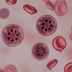
The images in the dataset were captured by placing a smartphone over a microscope to capture the Field of View (FOV) of the blood slide through the eyepiece of the microscope. Along with the image, the slide from which the image was captured, the stage micrometer readings of the microscope, and the objective lens settings were recorded, and a maximum of 40 images was captured from each slide.
This blood slide image dataset was curated to facilitate using Computer Vision techniques for quick and accurate diagnosis of malaria in low-resource settings. This dataset adds to existing malaria microscopy datasets and can be used to improve machine learning models to generalise to data collected in other communities like Uganda.
There are 2 747 images in the train and 1 178 in the test.
Access the images here: https://drive.google.com/file/d/16T40TdpaB8VXohm50SySREwrzbuPcJBC/view?usp=sharing
Join the largest network for
data scientists and AI builders